Capsular bag distension syndrome (CBDS) is an unusual complication of
cataract surgery. It is characterized by an accumulation of fluid between the posterior chamber intraocular lens (IOL) and the posterior capsule. Sometimes this fluid can be visualized easily at the slit lamp as a turbid liquid posterior to the IOL.
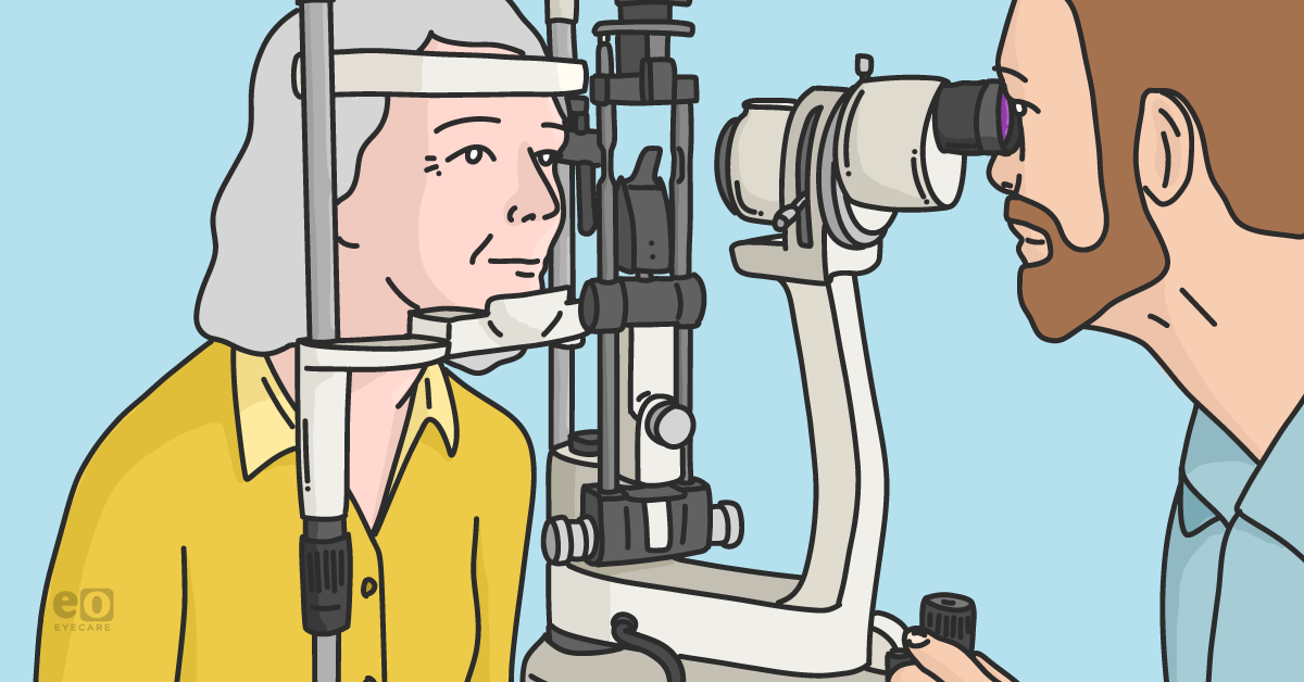
Other times, the fluid can be nearly invisible and is only appreciated with anterior segment imaging, such as ultrasound or
AS-OCT, where it appears as a hyperreflective diffuse opacity posterior to the PC-IOL.
Capsular Bag Distension Syndrome Overview
CBDS is a rare phenomenon. A retrospective chart review in South Korea found that out of 1,100 eyes, only eight cases of postoperative CBDS were found.1 That same study found that risk factors included an axial length over 25mm and the use of the Akreos Adapt 4 haptic PC-IOL.
Symptoms of CBDS
Symptoms of CBDS can vary, depending on the speed of onset and clarity of the fluid. Oftentimes, patients will have symptoms consistent with posterior capsular opacification (PCO), such as a progressive clouding of vision and glare.
2 If a patient presents with these symptoms without a significant PCO, there may be subtle CBDS at play. Anterior segment imaging can be helpful in these situations. Another common symptom is an
unexpected myopic shift.
2As fluid accumulates, the PC-IOL will be displaced anteriorly, inducing myopia. There have been reports of a hyperopic shift, thought to be due to a concave lens effect between the curvature of the posterior surface of the IOL and the posterior capsule.3
Beware, as this anterior movement can narrow the iridocorneal angle and shallow the anterior chamber, along with causing an
increase in intraocular pressure. However, there have been reports of a lack of a myopic shift due to a sufficiently fibrotic anterior capsule that resists anterior movement.
4Identifying types of CBDS
Miyaki et al have distinguished three types of CBDS based on time of onset.5
- Intraoperative CBDS occurs when balanced salt solution (BSS) becomes entrapped in the space posterior to the PC-IOL. This increases the risk of a posterior capsular tear.
- Early CBDS, which presents within 2 weeks of cataract surgery, is likely due to viscoelastic trapped behind the IOL. The viscoelastic can also create an osmotic gradient that causes the viscoelastic to swell and increase in volume. This can cause a myopic shift, narrow angle, shallow chamber, and elevated IOP. Early CBDS can be treated with a YAG capsulotomy.
- Late CBDS presentations, occurring months to years after cataract surgery, are thought to be due to lens epithelial cells that have been sequestered posterior to the IOL. These epithelial cells proliferate and produce a turbid fluid that fills the space between the IOL and the posterior capsule. Further evidence of this theory is that alpha-crystallin proteins have been found in aspirates of this fluid.6 Late CBDS can cause clouding of vision and reduction in visual acuity. It can be treated with a Yag capsulotomy.
Treatment options for CBDS
Treatment of CBDS typically involves a capsulotomy to allow the fluid to drain and thus clear the visual axis.
2 Most commonly, a
YAG capsulotomy is performed on the posterior capsule. However, if the posterior capsule cannot be visualized adequately, an anterior YAG capsulotomy can be performed. Intraocular pressure must be monitored carefully if an anterior YAG capsulotomy is performed because the sudden inrush of fluid in the anterior chamber can cause an acute IOP spike.
7Caution should be taken when performing a capsulotomy because there have been reports that the entrapped fluid may contain bacteria, such as Propionibacterium acnes.2 Releasing these bacteria into the eye can cause endophthalmitis.
To reduce the risk of
endophthalmitis, surgical aspiration can be performed. This procedure involves penetrating the anterior capsule, removing the fluid, and lavaging the space between the IOL and the posterior capsule. Injection of antibiotics follows.
8For a more complete removal of the material, a pars plana vitrectomy can be performed, followed by aspirating the fluid. Galvin et al argued that this may further reduce the risk of endophthalmitis.
9 However, it also exposes the patient to the risks inherent in a vitrectomy, such as
retinal detachment.
Case report
An 81-year-old Caucasian female presented as a new patient with a chief complaint of “cloudy” vision in her left eye. The patient felt that her vision began clouding approximately 2 months prior to presentation. Her ocular history was significant for uncomplicated,
bilateral cataract extraction 6 years ago by an outside ophthalmologist.
She felt that her symptoms were worse while driving and that
artificial tear usage had not improved her symptoms. Her medical history was significant for osteoarthritis, osteoporosis, and hypercholesterolemia, for which she was taking meloxicam, alendronate, and atorvastatin, respectively.
Corrected visual acuity was 20/30 in each eye, with glare acuity of 20/60 OD and 20/200 OS. Ocular motility was full, confrontation visual fields were full to finger counting, and pupils were equal, round, and reactive with no afferent pupillary defect. Intraocular pressures were 11mmHg OU via Goldmann applanation tonometry.
Anterior segment examination revealed
dermatochalasis and
meibomian gland dysfunction in both eyes, as well as clear PC-IOLs in both eyes. Of note, there was no significant posterior capsular opacification in either eye. However, a close examination of the left eye revealed a turbid fluid between the PC-IOL and the posterior capsule. Dilated
fundus examination revealed a cup-to-disc ratio of 0.4 OU with healthy-appearing optic nerves and no evidence of maculopathy.
A diagnosis of delayed capsular bag distension syndrome was made, and the patient underwent an uncomplicated YAG capsulotomy OS. At her 3 week post-operative visit, corrected acuity OS had improved to 20/25, and the patient reported a significant improvement in her vision. The patient will be followed closely for the development of endophthalmitis.
Conclusion
Capsular bag distension syndrome is a rare complication following uneventful cataract surgery. It can present any time after cataract surgery with symptoms initially suggestive of posterior capsular opacification. If a patient presents with symptoms of decreased visual acuity and glare/haze and no significant
posterior capsular opacification is found, capsular bag distension syndrome should be on the list of differentials.