

As eye doctors, one of the most frustrating and concerning situations we face is not being able to determine the reason for reduced visual acuity.
We have all been there.
It’s the end of the exam and despite our best efforts to examine the ocular structures of the eye and improve the patient’s vision with a thorough refraction, nothing adequately improves the patient’s vision or fully explains the reason for their reduced visual acuity.
In these moments, a simple formula can be followed to discover the etiology of vision loss or reduced visual acuity.
Because there are only a limited number of refractive structures in the eye that can obstruct light flow, one must look towards utilizing other tests closely examine some of the most common sources of reduced visual acuity.
Two of the most helpful diagnostic tools can be optical coherence tomography (OCT) and corneal topography.
Check out this in-depth guide on utilizing OCT for the treatment and management of retinal pathology.
These two simple tests can reveal the vast majority of unexplained causes in reduced visual acuity.
Many conditions of the macula and cornea are simply not visible with standard biomicroscopy examination. (Consider the difficulty of identifying early keratoconus, irregular astigmatism, macular pucker, vitreomacular adhesion, or early stage macular holes, etc).
Many conditions may not be visible without ancillary testing. If you do not have access to an OCT or Corneal Topographer, send the patient to an office that can perform these tests for you.
What happens if your OCT and Corneal Topography come back within normal limits?
After all primary structures of the eye that typically cause reduction in visual acuity (cornea, macula, lens) have been ruled out, then it is time to consider neurological cases.
When I say “neurological cases” I use that term very broadly to mean any disease of the optic nerve or brain. Testing for neurological conditions can be quite complicated; however, a simplified approach is to perform a few basic tests. First, study the optic nerve head for any pallor, do a red cap desaturation test, and carefully look for asymmetric pupil responses to light.
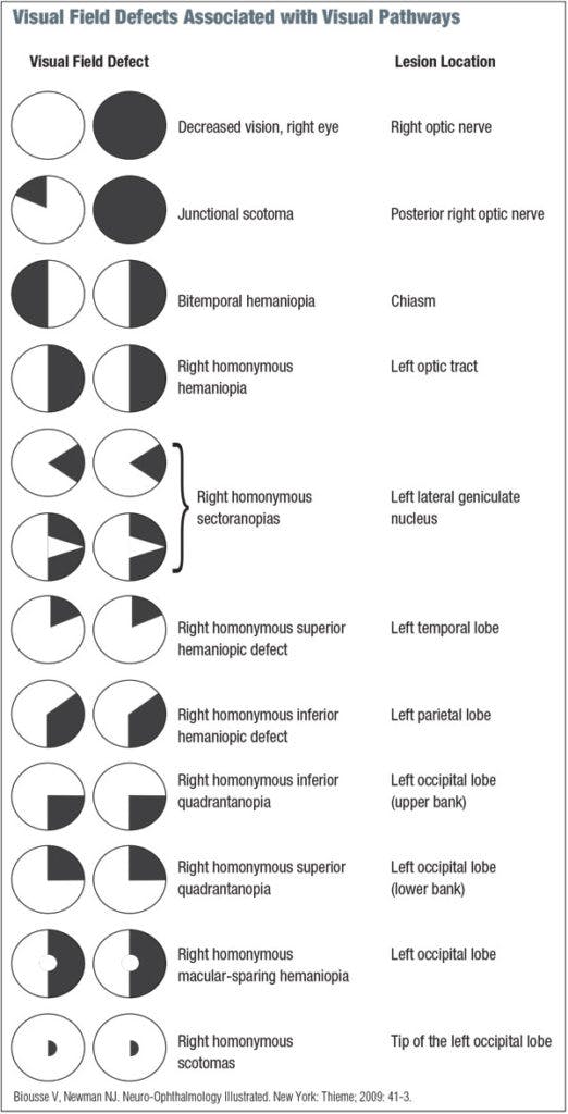
After those tests, order an OCT of the optic nerve and an automated visual field test. A visual field test is so important because the type of visual field defect will clue you in to the cause of the disease. Different neurological conditions exhibit different characteristic types of field loss.
Check out this fantastic overview of visual field defects associated with the visual pathways:

In summary, follow the simple formula of:
1) Topography of cornea and OCT of macula
2)Visual Field and OCT of Optic Nerve
These will help discover the vast majority of unexplained reduction in visual acuity. Part of the fun of being an eye doctor is searching for the hidden cause of vision reduction and then uncovering the diagnosis.
There are some “Zebras”/rare disease entities that will still not be discovered by this method, but those cases will be exceedingly rare. If you have done all the testing described above and you are still unsure of the diagnosis, then that is a good time to get a second opinion.
However, don’t be too quick to refer it out. If you push yourself a little bit and take the time to run the appropriate tests, you will be pleasantly surprised at what you are capable of and the patient will be grateful for not having to see another eye doctor.

